Day 1 :
Keynote Forum
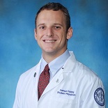
Samuel G. Eaddy
Nova Southeastern University College of Osteopathic Medicine, FL 33314, USA
Keynote: Dr
Time : 09:00 to 09:45

Biography:
Samuel Eaddy has completed his Master’s in Physiology and Biophysics from Georgetown University and is currently an OMS-II at Nova Southeastern Univeristy College of Osteopathic Medicine. He has conducted clinical research in various specialties since 2014, and is currently working with a team of residents and medical students to advance multiple orthopaedic studies at the Boward Health Medical Center, which is home to a reputed orthopaedic surgery residency program in South Florida.
Abstract:
Sacral dysmorphism (SD) is a congenital anomaly found in up to 54% of the population. This includes abnormalities within the lumbosacral joint and its surrounding structures, presenting increased risk in the surgical repair of posterior pelvic ring injuries. Iliosacral and transsacral screw fixation is greatly influenced by these anatomical variations, consequently altering surgical planning. A systematic review was performed with the following objectives: to determine the overall prevalence of SD; to summarize the implications of its anomalies in surgery; and to describe the altered safe zones that may be available to orthopaedic surgeons. Inclusion criteria included studies that reported the statistical prevalence of SD and their associated features. Data collected included patient demographics, prevalence of SD, quantification or classification of dysmorphic anatomy, and postoperative complications. Our analysis demonstrated a prevalence of 23% among 1,983 pelves in 11 studies. Among the seven known dysmorphic criteria, only three have been considered significant in the evaluation of screw placement. Approximately 95% of dysmorphic sacra can accept an S2 transsacral transiliac screw compared to 50% in normal counterparts. Additional evidence suggests viable fixation pathways in dysmorphic S3. These results led to the conclusion that SD is a relatively common condition that appears to present on a spectrum of severity, yet the variability in dysmorphic anatomy complicates the development of a universal solution to biomechanical fixation. Standard protocol suggests fixation of dysmorphic sacra at S2 when S1 is not viable, as lower sacral segments have been found to yield greater opportunity in this patient population.
- WEBINAR ON ORTHOPEDICS AND ARTHROPLASTY
Location: Online
Session Introduction
Khatri B
Central Institute of Orthopedics, VMMC And Safdarjung Hospital, New Delhi Delhi, India
Title: ROLE OF CT SCAN GUIDED BONE MARROW INJECTION IN POSTOPERATIVE CASES OF FRACTURE FIXATION WITH NONUNION IN LONG BONES
Biography:
Our Firm, B. Khatri & Co., provides wide range of services ranging from international tax, domestic tax (both direct and indirect), transfer pricing, expatriate tax and regulatory matters, representation, assisting in business reorganizations, tax due diligence, etc. We also provide assurance, accounting, payroll, valuation, incorporation, incubation, virtual office services among others. Since the introduction of GST, we have been involved in assisting client.
Abstract:
Fracture non-union is a relatively common problem faced by orthopedic surgeons after operative fracture fixation. Percutaneous Bone marrow injection given at non-union site has successfully shown union with significant low morbidity as compared to traditional open bone grafting. The purpose of our study was to use CT scan guidance to accurately locate the exact site of non-union, deliver an effective shot of autologous bone marrow injection and assess union time. Method: Under strict aseptic precautions bone marrow aspiration was done from posterior iliac crest using core biopsy needle in a heparinized syringe. Under CT guidance, multiple punctures were done over sclerosed non united fracture ends followed by percutaneous injection of bone marrow to stimulate union. All patients were followed up with serial radiographs and clinically with appropriate scoring systems. Results: 30 patients were selected for hospital based interventional based study with average time after operative fracture fixation of 9.5 months. After bone marrow injection, Follow-up period ranged from 6 to 8 months with average union time of 4 months and complete union in 24 out of 30 patients (80%). Failure was reported in six patients (20%).Conclusion: CT SCAN guided bone marrow injection is an effective method for treatment of delayed or non-union fractures with low complications. It has significant low rates of morbidity as compared to autologous bone grafting procedures and it reduces patient’s expenditure, hospital stay and gives good functional results. It can be tried for non-union before going directly to gold standard re-fixation and autologous bone graft.
Alexander Frost-Younger
Royal Victoria Infirmary, Newcastle upon Tyne, United Kingdom
Title: LENGTH OF STAY COMPARISON IN PATIENTS REQUIRING SURGICAL INTERVENTION FOR WRIST FRACTURE: A SMALL STUDY FROM A TRAUMA CENTRE

Biography:
I attended Newcastle University and graduated in 2016 with a Bachelor of Medicine and Surgery. I hold a GMC Registration with 'Full License to practice' and completed my Foundation Programme Training in the Northern Deanery 2016-2018. During my medical education, I have developed an interest in Emergency/Acute Medicine, Surgery and Radiology. I will be applying for specialty registrar training in Surgery this year.
Abstract:
Authors: Dr Alexander Frost-Younger. Royal Victoria Infirmary, Newcastle upon Tyne, United Kingdom NE1 4LP
Background: Isolated wrist fractures are one of the most common fractures in both children and adults. In 2008 the overall fracture incidence for the UK population was calculated at 3.6%(1). However, studies comparing patient length of stay (L.O.S) for fracture surgery are sparse.
Method: A small retrospective study examining 90 patients with isolated wrist fractures requiring surgical intervention between Sept 2018-Sept 2019. Exclusion criteria included multiple fractures, polytrauma and concomitant medical conditions on admission. Patient Demographics and admission dates (Winter/Non-winter and Weekday/Weekend) were compared by calculated L.O.S averages.
Results: Patients >60 (n=2.1), patients requiring ORIF (n=2.0) ASA Grade >1 (n=1.9) had the highest average L.O.S. Seasonal and weekly variation was noted, with patients having a higher L.O.S if they were admitted during Winter (n=1.7) or over the weekend (n=1.6), in comparison to Non-Winter and Weekday admissions.
Arpit Patel
Royal Free Hospital, London, United Kingdom
Title: Septic arthritis secondary to PVL toxin producing staphylococcus aureus
Biography:
Mr Arpit Bakulash Patel MBCHB, BSC(Hons), MRCS (England), PGCert (Medical Education) Core Surgical Trainee, London Deanery
Abstract:
Introduction
Panton-Valentine Leukocidin (PVL) toxin producing strains of staphylococcus aureus are known to cause severe skin and soft tissue infections. We present a case report of a patient who suffered from septic arthritis and overwhelming sepsis from proven PVL toxin secreting staphylococcus aureus.
Case Study
A 13 year old boy presented to the Emergency Department with a one day history of right sided hip pain and an inability to weight bear, one week after incision and drainage of a left sided buttock abscess. He was systemically unwell with raised inflammatory markers and continued to deteriorate despite Co-Amoxiclav and washout of the hip. Post-operatively, ongoing pain led to an MRI that showed worsening right hip septic arthritis with extensive inflammatory changes. A change of antibiotics to Rifampicin and Trimethoprim following PVL isolation and second washout led to improvement.
Discussion
The literature supports that beta-lactam antibiotics increase the expression of staphylococcus aureus PVL toxin by activating its transcription leading to a greater systemic inflammatory response. Patients usually present with minor skin infections however rarely, invasive infections can occur including septic arthritis. An MDT effort to achieve early surgical management with tailored antibiotics can improve outcome as the use of penicillin can be detrimental.
